Summer Sale
Buy 1 pair: 30% off frame
Get 2nd pair: 50% off frame and lenses
Disclaimer: Sale ends August 31 2025. Some restrictions apply.
At York Eye Care Clinic our number one priority is to perform the most comprehensive eye examination providing you the best possible prescription for eyeglasses or contact lenses and to diagnose and treat ocular diseases. Our vision care service also includes refractive laser surgery consultation co-management and myopia management.
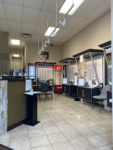
It is our mission to provide the most thorough care to each and every patient. We strive to always provide you with the best service, most current information and education and the best products on the market. For your convenience, our new and modern optical dispensary carries a large selection of designer and value eyewear. Our staff will provide exceptional customer care to meet and exceed your expectations.
We pride ourselves on your satisfaction.
We are conveniently located in the Aurora, Newmarket area at 302 Wellington Street East in the Tim Horton’s plaza across from St. Maximilian Kolbe Catholic High School.
You can reach us at 905-751-0007


Seniors 65 years and older and those 19 and under, are covered by OHIP for a routine eye examination provided by an optometrist once every 12 months plus any follow-up assessments that may be required.
Patients aged 20 to 64 years who have any of the following medical conditions can go to their optometrist and receive an OHIP insured eye examination once every 12 months : diabetes mellitus, glaucoma, cataract, retinal disease, amblyopia, visual field defects, corneal disease, strabismus, recurrent uveitis or optic pathway disease.
Routine eye examinations for patients aged 20 to 64, are not covered by OHIP.
Direct billing is a service provided by our clinic that makes it easy to submit insurance claims electronically on your behalf. This saves you time and eliminates the hassle of dealing with your insurance provider, just like when you go to the pharmacy or the dentist, which can reduce your out-of-pocket expenses.
Do you offer direct billing in your Clinic?
Yes, we offer direct billing with most insurance companies. You will need to bring your health insurance card featuring your member identification and policy number.
Even if your insurance company is listed below, your specific plan may not allow for online submissions. Please verify with your insurance provider prior to your appointment.
Please note, with some insurance companies the payment goes directly to the insured member, in that case we can still electronically submit a claim on your behalf.
Will my insurance cover my eye exam and glasses/contacts?
Please contact your insurance provider to determine your eligibility for your vision coverage prior to your appointment.
Insurance We Accept
For your convenience, we accept most health insurance providers.
Click here to see a list of insurance companies that are accepted at our location.
If your insurance company is not listed here, please contact our office as soon as possible to see if we can help you.
We hope you will consider York Eye Care clinic for your Optometric needs.
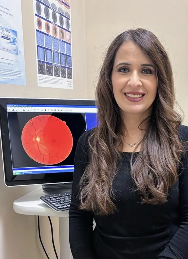
A comprehensive eye exam uses a wide range of tests and procedures to test your vision and ocular health. Diagnostic tests such as autorefraction or retinal imaging will be performed prior to your eye exam. This gives the doctor a chance to review the results before a thorough examination is started.
In the exam room a detailed history of your medical and ocular health is taken. So please bring in a list of all your medications as systemic conditions may have ocular impact.
Visual acuity:
To determine the sharpness of your vision, you will be asked to read an eye chart from a computer. The letters will progressively get smaller as you read them out loud until you no longer can see them.
Color Vision Test:
You will be asked to read out numbers from a book to determine if you are color deficient. 10% of all males and 2% of females are color blind.
Stereoscopic vision test:
This test determines how well both eyes work together. You will be given 3-D glasses for this test and will be asked to identify the raised objects from a book. Stereoscopic vision test is important in identifying diseases such as Amblyopia, Strabismus, Suppression and stereopsis.
Cove test:
This is an objective determination of the presence and the amount of ocular deviation. The doctor will cover and uncover each eye with a paddle while you are looking at a target at a close range or at a distance. The misaligned eye will deviate inwards or outwards and the amount of it can be measured using a prism bar.
Refraction:
To determine your exact prescription, the doctor will use a phoropter by asking you to respond to questions “which is better, one or two?” while flipping back and forth between different lenses. Remember to relax and respond what you see as there are no wrong answers.
Slit lamp Exam:
A slit lamp which is a highly magnified device is used to examine the health of the eye. Eye condition such as dry eye, corneal dystrophy, cataract, uveitis and abnormality of the iris can be seen during this test. Furthermore, the doctor can use a special 90D lens to view the retina. Conditions such as glaucoma, macular degeneration, diabetic retinopathy can be diagnosed.
Goldman Tonometry:
Eye pressure are measured by placing an anesthetic drop and a flurescene dye in the eyes. Then a tool called tonometer is used to measure the pressure of the eyes. The tonometer is highly accurate and is the “gold standard” for glaucoma. Patients should not hold their breath during the exam and can breath slowly through their nose.
Retinal Examination:
To assess the health of the retina, the doctor will put drops in the eyes to dilate them. This will provide much wider view of the retina and conditions such as retinal holes, tears and detachment can be detected using a binocular indirect ophthalmoscope (BIO).
The Entire exam can take anywhere from 20 minutes to one hour.


If your insurance company is not listed, please contact the clinic.
To make an inquiry or to book an appointment, please fill and submit the form below.