Retinal Imaging & Visual Field Testing

What is Retinal Imaging?
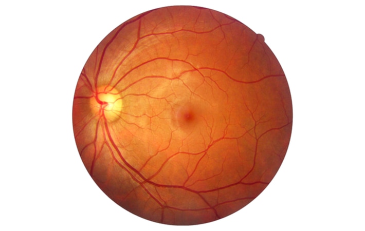
Retinal imaging is a non-invasive, diagnostic tool that produces colored images of your retina, optic nerve, and blood vessels in the back of your eye. This will help doctors assess the health of your retina and helps them to detect and manage such eye and health conditions as diabetes, glaucoma, and macular degeneration. Finding retinal disorders as early as possible is critical to potentially preventing serious disease progression and even vision loss.
Images can be compared side-by-side, year after year, to discover even subtle changes and help monitor your health. They allow your doctor to explain treatment more thoroughly as you can review the images together, which ensures a certain level of precision to your routine eye exam.
Do I need a Retinal Imaging?
Incorporating retinal imaging into an eye exam may be beneficial to establish a baseline of your retina. Subtle changes of the retina can be determined earlier by comparing retinal images that otherwise may not be able to be detected by looking with the naked eye.
The following are some of the conditions that can be seen on a retinal imaging:
Age-related Macular Degeneration
Macular degeneration is usually signified by leaking of fluid or bleeding in the back of the eye. This causes central vision loss.
Diabetic Retinopathy
Diabetes can cause changes in the blood vessels of the retina, like swelling and leakage or the creation of new blood vessels. Blindness can result without early detection.
Hypertension (High Blood Pressure) / Hypercholesterolemia (high chlestrol)
Signs of high blood pressure and cholesterol often appear in the eye. Indicators can include narrowing of the blood vessels, exudates (spots on the retina), or bleeding in the back of the eye.
Glaucoma
Pressure against the optic nerve and compression of the eye’s blood vessels may indicate glaucoma. This disease may cause permanent and irreversible vision loss.
Retinal Detachment
Retinas can lift or pull away from the wall of the eye. If not properly treated, this can cause permanent vision loss.
Cancer
A dark spot at the back of the eye may signal a melanoma, which can grow unnoticed within the retina. If caught early, melanomas can be treated before they cause serious damage and spread to other areas of the body through the bloodstream.
What should I expect during a retinal imaging test?
This test is as easy as taking a picture. You will be asked to place your chin and forehead on supportive rests to help you keep your head still. Next, you will open your eyes as wide as possible and stare at an object straight ahead. A camera flash will be seen when the photograph is taken, capturing high definition images of your retina and optic nerve.
The images are displayed on a computer screen for your eye doctor to review with you.
Is retinal imaging safe?
Retinal imaging is a non-invasive & completely safe method of obtaining pictures of the back of the eye. You can rest assured that this is a relatively quick and painless test.
The entire process takes around 5 minutes.
However, never hesitate to speak with your eye doctor if you have any concerns about the test, or your ocular health.
Visual Field Testing
What is a visual field test?
The visual field test is a way to measure your entire visual field. This area includes all the range of what you can see above, below, and on either side of you while your gaze is fixed on a central point. A visual field test enables your eye doctor to measure the range of your peripheral vision, identify any abnormalities in your visual field, such as blind spots (scotoma), or vision loss that may have occurred acutely such a stroke or over time such as glaucoma.
Why is the visual field test important?
Visual field defects have a number of causes, including disorders that don’t originate in the eye. You may have a healthy eye but have a visual field defect, which might indicate a problem in part of the brain that deals with vision. It is also important in diagnosis of retinal conditions and retinal nerve damage. Furthermore, visual field tests are used to assess how vision may be limited by eyelid problems such as ptosis (droopy eyelids).
Your doctor may use the information from the visual field result to diagnose:
- macular degeneration
- galucoma
- optic glioma
- brain tumor
- stroke
- multiple sclerosis
- temporal arteritis
- central nervous system disorders
- pituitary gland disorders
- high blood pressure
Your doctor may order further eye tests to diagnose a problem. If an eye problem is not indicated as the cause of vision problems, your doctor may send you for a physical examination and blood tests.
Do I need a visual field test?
Many ocular conditions can increase your risk of retinal nerve damage or vision loss. The visual field test can help the doctor find early signs of diseases like glaucoma that damage vision gradually. Some people with glaucoma do not notice any problems with their vision, but the visual field test shows that peripheral vision is being lost.
Your eye doctor will also test your visual field if you have been diagnosed with any of the following health conditions, as these conditions can lead to sight-threatening ocular conditions:
- Hyperthyroidism
- Multiple sclerosis
- Pituitary gland disorders
- Stroke
- Brain tumor
- Diabetes
- Hypertension
If you notice any changes in your vision, schedule an eye exam as soon as possible to rule out any ocular conditions that can lead to vision loss.
A visual field test can facilitate early detection of vision loss, enabling earlier treatment and increased optimal results.
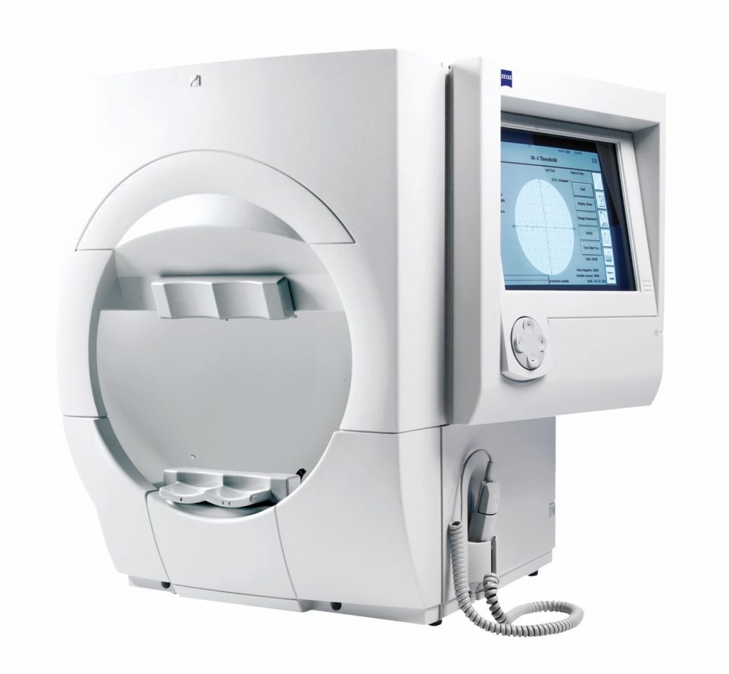
Picture of the machine is shown below;
What should I expect during a visual field test?
During this test, you will sit in front of a bowl-shaped device, called a perimeter. Each eye will be tested separately. The eye being tested will look through a lens containing your optical prescription to ensure that you can see well.
Your doctor will instruct you to keep your eye on the center target throughout the test. Small oval lights begin to flash in different areas of the bowl, some dimmer than others. The patient presses a button to indicate when lights are seen and the instrument records which were not seen. Any lights that you did not see will be tallied at the end of the test, and provided in a print out for easy reading.
Part of the test is how well a person can see really dim lights to determine how well you did at the extreme of your ability. This will determine the visual threshold, or the lights you cannot see some of the time. In fact, during a field test the machine is going to show you lights 1/3 of the time that it knows you can’t see — so don’t worry if there is a point in the test where you haven’t pressed the button for a few seconds, this is normal.
Let’s Get Started
To make an inquiry or to book an appointment, please fill and submit the form below.